Wednesday, November 19, 2014

Week 4

This week I observed many of the same organisms as previous weeks.  The largest and smallest organisms increased in number and the medium sized organisms decreased in number since the last observation. Diatoms seemed to get the biggest increase in number and size this week.

Below are diatoms of genus Pinnularia (Canter-Lund and Lund, 1995 p.118 Fig. 207).  The biggest one (Diatom A) was seen moving by moving water through the tube like body and propelling itself left or right.

Many were idle and floating by themselves, some of which (pictured below) were stacked for better structural protection from predators.


 This nematode of genus Tylenchorhynchus (below) was the only new animal spotted this week (Allen 1995).  It seemed to love to stay shielded in a layer of dirt, but was faster moving than the Duro annelid spotted last week (Covich and Thorp 2009, p.399 Fig12.5).
 This protist of genus Stentor was spotted near the bottom of the tank (MicrobeWiki, 2014).  It uses a filter feeding technique to bring food particles to it.  This two headed Stentor was attached to a decaying stem.
 This un-identified rotifer was spotted holding onto and spinning around the photosynthetic plant Amblestegium varium (McFarland, 2014).


Monday, November 10, 2014

Week 3

Over the past week the number of both large organisms and small organisms increased.  New species of each were spotted.  There were many stationary diatoms and small dead organisms.
This unidentified protist showed up near the leaves of my plant to eat some algae growing there.  The front end was covered in cilia.

Six members of genus Actinosphaerium were spotted.  The spike protruding from the mucilaginous layer around the cell are known as spicules.  Five of them were connected by specialized tubing (Patterson 2013, p.169 Fig.395)

One shelled amoeba of genus Centropyxis was spotted near the middle of the tank not moving (Patterson 2013,p.95 Fig.184).

One member of Gastrotricha genus Chaetonotus was spotted swimming in the lower middle of the tank (Pennak 1989, p.165 Fig.7).

I spotted the tail end and oxygen harvesting gills of an immature annelid of genus Dero hiding among the muck at the bottom of the tank (Covich and Thorp 2009, p.399 Fig12.5).

Friday, October 31, 2014

Week 2

This week my microaquarium was teeming with life.  On Friday October 24, 2014 "ONE" Beta Food Pellet was inserted into each microaquarium. On your blog posting for this week include date the food pellet was added along with the following information: "Atison's Betta Food" made by Ocean Nutrition, Aqua Pet Americas, 3528 West 500 South, Salt Lake City, UT 84104. Ingredients: Fish meal, wheat flower, soy meal, krill meal, minerals, vitamins and preservatives. Analysis: Crude Protein 36%; Crude fat 4.5%; Crude Fiber 3.5%; Moisture 8% and Ash 15% (McFarland, 2014).  Anywhere I looked through the microscope there were tiny organisms eating and moving around.  The food pellet seemed to increase the amount of organisms tenfold.

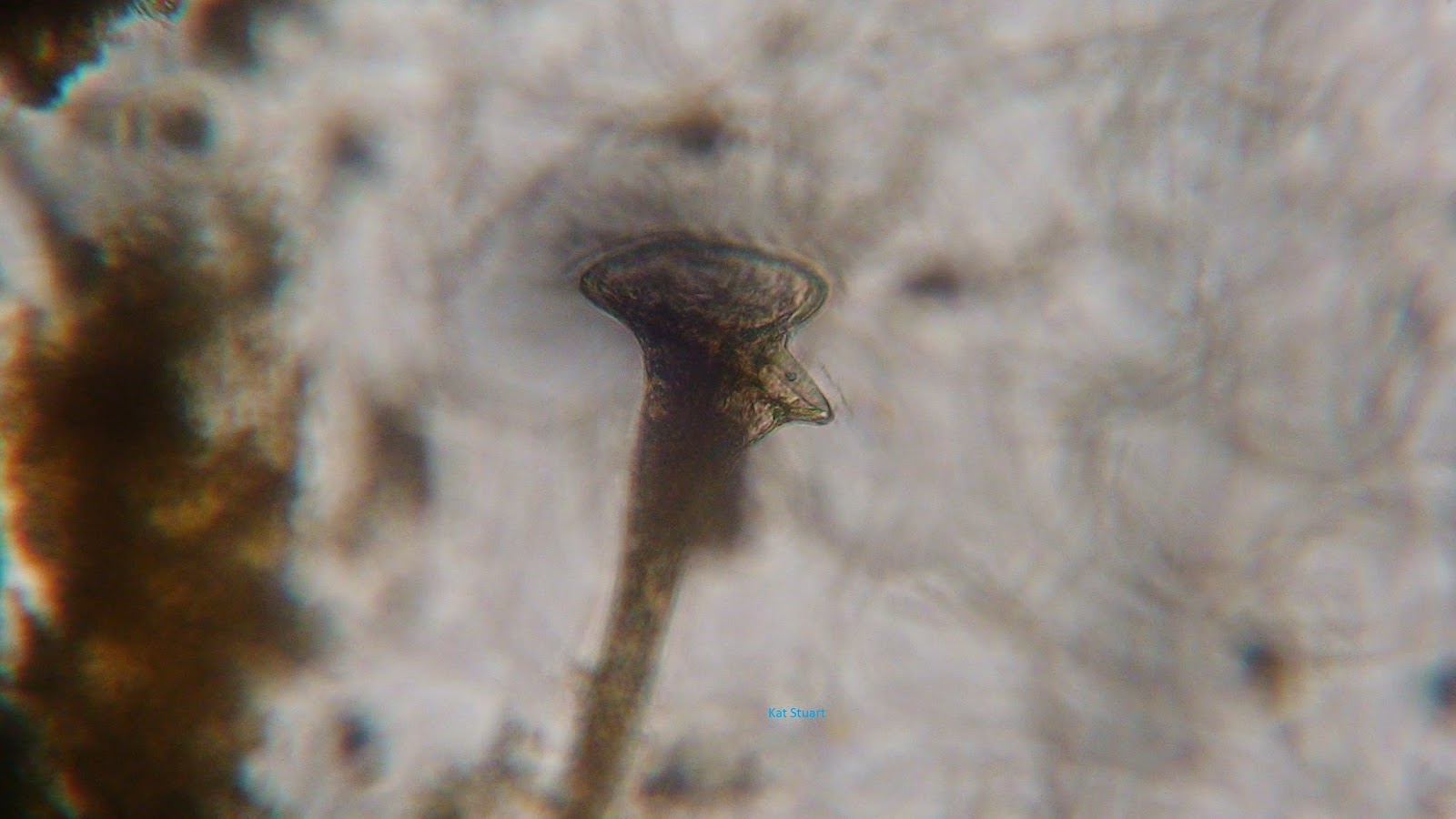
I saw this microorganism moving along a stem about midway through the tank.  Only one was observed. 


This amazing Epistylis (Patterson 2013) was spinning the circular portion in the lower left corner to draw particles into it to capture food.  This organism was tethered to a steam near the middle of the tank and was seen nearby a few other of its kin.
This organism also was tethered to a stem in the middle of the microaquarium and moved the bottom portion of its body to bring particles to it.  It was seen alongside two other organisms of the same kind.
This unsegmented worm-like organism was seen near the bottom of the tank eating whatever smaller organisms came near it.  The digestion process was very interesting to watch.  The ingested organisms would be pushed to the back of the large chamber in the middle of picture and then would be pushed past a sphincter into another digesting chamber that was mostly obscured from view in the soil. Only one was seen.
This Philodina (Pennak 1989) was found near the middle of the tank spinning around the water column while the large end of it pulled particles into it. A few were observed near each other with nothing else in close view.  This suggests the Philodina are successful competitors in my microaquarium. 

Sunday, October 26, 2014

Week 1

The organisms in the water collected from Spring Creek in Dean's Woods N35 55.274 W083 56.888 848 ft on 10/12/2014 have become more plentiful (McFarland 2014). 
Many of the creatures observed were close to the dirt layer or found directly around the living plant tissue.  Many other moving organisms were observed but were too small to take a picture of.
 1
Euchlanis sp. is a member of Rotifera (Pennak 1989) and was found scooting around the water in low density debris near the middle of the water.  Only one was observed.


 2
Litonotus sp. (Patterson 2013) was found reaching its way across and around the plant material.  Only one was observed.


3
This unID'd rotifer(?) was spotted near the substrate at the bottom of the tank.  Only one was spotted.

4
This unID'd rotifer(?) was spotted near the substrate at the bottom of the tank.  Only one was spotted.

5
A few of these unID'd Protists(?) were observed


6
Anisonema sp. has one larger recurrent flagellum that drags (pictured) and an anterior flagellum that beats normally. (Patternson 2013) One Anisonema was spotted near plant tissue. 

Monday, October 20, 2014

Bibliography


Allen. 1995. [Internet] The Cilicate Resource Archive. [cited 11/19/2014]. Available from http://www.uoguelph.ca/~ciliates/repgenera/stentor.html
Canter-Lund H, Lund J. 1995.  Freshwater Algae: their microscopic world explored. 1st ed. UK: Biopress Limited.
Cook R, McFarland K. 2014. General Botany 111 Lab Manual. 16th ed.  TN: University of Tennessee. 
Covich A, Thorp J. 2009.  Ecology and Classification of North American Freshwater Invertebrates.  3rd ed.  MA: Academic Press. 
McFarland, Kenneth [Internet] Botany 111 Fall 2014. [cited 10/22/14]. Available from http://botany1112014.blogspot.com
MicrobeWiki [Internet] Stentor. [cited 11/19/2014]. Available from https://microbewiki.kenyon.edu/index.php/Stentor
Patterson DJ, Hedley S.  2013.  Free-Living Freshwater Protozoa.  5th ed.  London:  Manson Publishing Ltd.
Pennak R. 1989.  Fresh-Water Invertebrates of the United States.  3rd ed.  USA: John Wiley & Sons.

Week 0


Microaquarium setup and Observations Week 0!

Today I set up my microaquarium complete with plants, dirt, and local water.  The microaquarium is made of two sheets of glass, a base, a stand, and a lid fixed with BlueStik.  I used water and dirt from  Spring Creek off Woodson Dr in Dean's Woods Road frontage., Knox Co. Tennessee. Partial shade exposure. Tennessee River water Shed N35 55.274 W083 56.888 848 ft 10/12/2014  (McFarland, 2014).  The water was collected from the bottom of the pool, the surface, and just over the sunken leaf litter.  I added clippings of two plants: one carnivorous plant (Utricularia gibba L. Original material from south shore of Spain Lake, Camp Bella Air Rd. East of Sparta Tn. in White Co. and grown in water tanks outside of greenhouse at Hesler Biology Building. University of Tennessee. Knox Co. Knoxville TN. 10/12/2014) and one solely photosynthetic plant (Amblestegium varium  Lindberg. A moss collected from Natural spring at Carters Mill Park, Carter Mill Road, Knox Co. TN. Partial shade exposure) (McFarland, 2014).  Stickers identifying my class seating position, lab time, and initials were added to the side of the aquarium.
After completing the microaquarium setup I oriented it horizontally under the microscope lens to observe the inhabitants.  I saw two types of microorganisms: one round organism using a spinning motion to move and pick away at small particles suspended in the water, and one eliptical shaped organism using a "shrink and push" style of movement to jet around the aquarium.  The organisms not preoccupied with feeding seemed shy of the light and tended to move away from the optical lens to hide in shadows.